Understanding Your Pathology Report

What is a pathology report?
A pathology report is a medical report that describes the characteristics of a tissue or blood specimen that has been removed from your body. The specimen is analyzed by a pathologist, who writes a report of their findings for the doctor who has ordered the report and/or performed the procedure.
Your pathology report contains important information about your specific colorectal cancer that helps guide decisions about your treatment. The report provides a definitive diagnosis as well as the stage of the cancer.
What will be in my pathology report?
Most pathology reports include these sections:
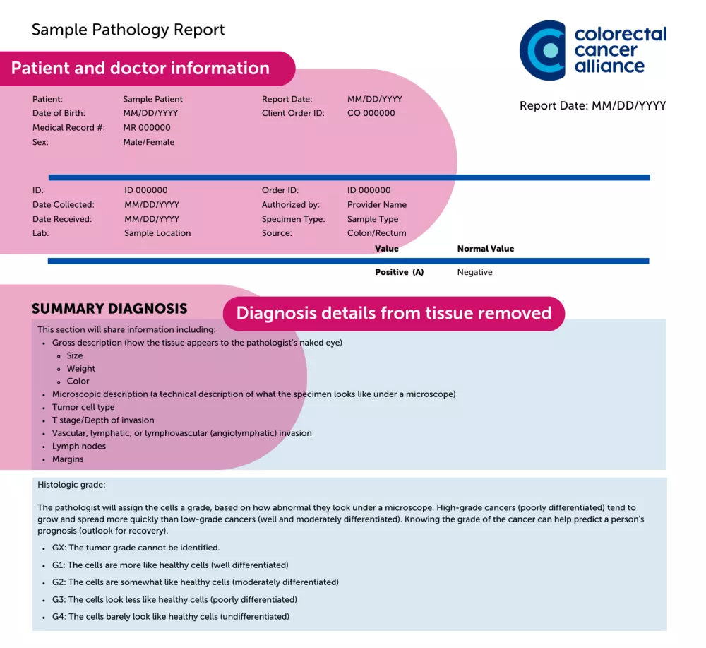
- Demographic information: Your name, birthdate, medical record number, presumed diagnosis, and the ordering doctor.
- Specimen type: From which part of the colon or rectum the specimen was taken.
- The type of procedure used to take the specimen, such as a biopsy or a specific type of surgery.
- Gross description: How the tissue appears to the pathologist’s naked eye, including size, weight, and color.
- Microscopic description: A technical description of what the specimen looks like under a microscope.
- Histologic grade: The pathologist will assign the cells a grade, based on how abnormal they look under a microscope. High-grade cancers (poorly differentiated) tend to grow and spread more quickly than low-grade cancers (well and moderately differentiated). Knowing the grade of the cancer can help predict a person's prognosis (outlook for recovery).
Colorectal cancer cells are assigned the following grades:
- GX: The tumor grade cannot be identified.
- G1: The cells are more like healthy cells (well differentiated)
- G2: The cells are somewhat like healthy cells (moderately differentiated)
- G3: The cells look less like healthy cells (poorly differentiated)
- G4: The cells barely look like healthy cells (undifferentiated)
- Tumor cell type: The type of cancer cells that are found in the tumor. There are several types of colorectal cancer. Adenocarcinoma is the most common type.
- T stage/Depth of invasion: This indicates the size of the tumor and how far it has invaded (spread) into the colon wall. Cancer that has not invaded the surrounding tissues is called in situ. A cancer that has penetrated the surrounding tissues is called invasive.
T (tumor) stage is classified as:
- Tx: The tumor cannot be measured.
- T0: There is no evidence of a tumor.
- T1s: The cancer cells are found only in superficial tissue (in situ)
- T1, T2, T3, or T4: Describes the size of the tumor based on size and depth of spread to surrounding areas.
- Vascular, lymphatic, or lymphovascular (angiolymphatic) invasion: When the pathologist examines the specimen and nearby tissue, they determine if the cancer has grown into the small blood vessels and/or lymph vessels of the colon or rectum. If it has, there is a higher chance that it could have spread outside the colon or rectum. This is not the same as cancer cells that are found in the lymph nodes.
- Lymph nodes: Cancer cells can travel through the lymph system to spread to other parts of the body. During colorectal cancer surgery, several lymph nodes are removed to be examined by the pathologist. The pathology report will state the number of lymph nodes that were examined and how many that had cancer cells in them. A lymph node is called “positive” when it contains cancer and “negative” when it does not.
- Margins: During surgery to remove cancer, the surgeon removes the tumor and some surrounding tissue. The pathologist then examines the edges (margins) of the specimen to see if there are any remaining cancer cells. A “positive” or “involved” margin means cancer cells were found in the edges.
- Diagnosis: In this section, the pathologist gives a definitive diagnosis based on the information above. If cancer has been found, the diagnosis typically includes:
- Type of cancer
- Tumor grade
- Lymph node status
- Margin status
- T stage/depth of invasion
- Stage of cancer

Questions to ask your care team
There are important details you’ll need to understand about your pathology report. Here are some good questions to ask your provider:
- What type of cancer do I have?
- How big is the tumor?
- What is the stage of the cancer?
- Is it a fast growing or aggressive cancer?
- Were the margins clean?
- Was cancer found in any lymph nodes?
- Do any tests need to be done again?
- Will I need any further surgery?
- How can I get a copy of my report?

What’s the difference between a pathology report and a biomarker test report?
A pathology report provides a diagnosis based on a pathologist’s examination of a tissue or blood sample, such as a biopsy of the patient’s tumor or blood sample. This diagnosis is made by evaluating laboratory tests, cells, tissues, and organs in the body.
A biomarker testing report provides very specific information about the type of cancer. Biomarker testing results are important because they can predict which treatments will be most effective and which ones will not.